
JoVE in Action is a series of blog posts highlighting how STEM educators around the world have used JoVE to support their remote teaching efforts. We hope these stories will be useful for instructors looking for effective ways to deliver their science and lab courses online or in hybrid formats.
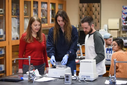
Dr. Mary Morrison is an Assistant Professor of Biology at Lycoming College. When her institution moved to remote instruction in mid-March, Dr. Morrison had to find a way to transform her upper-level Immunology course and its lab component into a virtual format. Because the course required students to submit two long lab reports based on raw data, she also had to create a virtual lab exercise that would enable students to fulfill this requirement.

To achieve this, Dr. Morrison first asked her students to watch JoVE Education videos on immunocytochemistry and immunofluorescence microscopy. “I jumped at this resource as the next best thing to teaching the students these techniques in a wetlab setting,” she said. She also created some questions based on these topics, and asked students to take notes as they watched the videos.
Then, she had a videoconference session with her students to ensure that they had understood the key concepts and to help them integrate the methods demonstrated in the two videos. She also suggests that educators could watch videos with students, via a videoconferencing platform, to answer students’ questions as they come up.
For the lab report, Dr. Morrison provided students with the following images, obtained in a previous semester’s Immunology course:
- Mouse cerebellar cultures stained with anti-calbindin D28k antibody with a Cy3 secondary antibody to show Purkinje neurons
- Mouse cerebellar cultures stained with anti-GFAP with an Alexa 488 secondary antibody to show glial cells
- A Differential Interference Contrast (DIC) image to show all the cells in the culture
- A no-primary-antibody control image
Using these images, and what they had learned about the relevant experimental procedures from JoVE videos, students were able to answer the questions drafted by Dr. Morrison and write a long-format lab report – despite no access to the wet lab.
Dr. Morrison notes that this method of teaching a lab course remotely is applicable to other science courses as well. “This virtual exercise could be used for advanced Immunology, Cell Biology, or Neuroscience courses,” she suggested. “It can be used with any set of fluorescent microscope images the instructor has available that are applicable to other areas of study.”
If you would like to learn how to effectively incorporate JoVE video resources into your hybrid, remote, or in-person science course, request a free consultation from our Customer Success team.