One of our editors highlights JoVE Video Articles which demonstrate the advantages of synchrotron-based X-ray techniques and their applications.
Synchrotron radiation facilities produce bright X-rays that can be applied by researchers in a wide variety of fields, including materials science, geoscience, and biology. These facilities can be used to answer scientific questions that cannot be addressed by benchtop techniques. While there are several advantages to synchrotron-based techniques because of the high intensity of the X-ray beam, the major limitation to synchrotron work is availability and access. With a limited number of these facilities around the world, researchers frequently must apply for time to use a technique at a remote lab and then travel to carry out their experiments.
JoVE video articles can provide researchers with a glimpse into synchrotron facilities without having to leave their benches. In this post, I’ll highlight some videos showing the advantages of synchrotron-based X-ray techniques and how to prepare for experiments at a synchrotron facility.
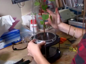
One advantage of synchrotron-based X-ray techniques is they are typically non-destructive. Unlike benchtop X-ray techniques where samples need to be placed under vacuum, synchrotron-based techniques can measure samples with minimal sample manipulation. For example, plant and mineral samples do not need to be dehydrated which can change their chemical or physical structure. In this JoVE video article, researchers demonstrate their methods for imaging the xylem networks in the tissues of living plants using high-resolution computed tomography.
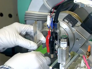
Another major advantage of synchrotron-based techniques is that data can be acquired rapidly. This allows for time-resolved data collection that can provide insight into processes in real time. In this JoVE video article, researchers at Lawrence Berkeley National Laboratory demonstrate how to conduct an in situ experiment to characterize how Li-ion battery electrodes change structurally during battery operation. Time-resolved X-ray diffraction patterns are collected to look for structural changes in the materials while undergoing battery charge and discharge.
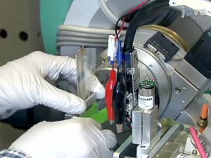
In another example of rapid data acquisition, researchers at Louisiana State University demonstrate how to use a lab-on-a-chip device to observe reaction intermediates in this time-resolved kinetic study. Researchers examining the synthesis of gold nanoparticles place a millifluidic device directly in front of the x-ray beam and collect x-ray absorption spectra during the course of the reaction.
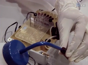
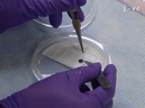
Proper sample preparation before arriving at a synchrotron facility is important to save time and ensure the success of an experiment. In this JoVE video article, researchers show how to prepare samples by chemically fixing cells on the appropriate sample holder. This process can be tricky because cells need to be grown onto a fragile sample holder and they can detach in transport if not done correctly. Following careful sample preparation in the laboratory, researchers are able to examine the distribution and quantity of elements in mammalian cells using X-ray fluorescence mapping.
These are just a few examples of the synchrotron-based methods published in JoVE. Since synchrotron-based radiation is not available to researchers in their labs, it can be difficult to become familiar with these techniques. Video articles can provide researchers who are new to synchrotron work with ideas on how to apply these advanced techniques to their research, what to expect when they arrive at a synchrotron facility, and how to prepare for a successful experiment.
